
Mar 20, 2020
 SIMFER – Italian Society of Physical and Rehabilitation Medicine has activated a teleconsultation service aimed at recently discharged stroke patients and their families who should continue rehabilitation treatments.
A.L.I.Ce OdV (Italian Stroke Association) spreads this new important free remote support and consultancy service offered by SIMFER Italian Society of Physical and Rehabilitation Medicine.
In consideration of the difficulties that many patients find in accessing physiatric visits and rehabilitation treatments, due to the limitations imposed by the current situation, SIMFER has activated (in Italy) in collaboration with A.L.I.Ce Ferrara odv a telemedicine-rehabilitation service, a sort of “virtual clinic”, made available in totally free form, which makes use of a selected group of physiatrists, able to offer indications and information support relating to the needs of people with disabling conditions of different origin.
The patient or caregiver who needs it, can write an email to SIMFER ITALY and will be contacted as soon as possible to carry out a teleconsultation with one of the SIMFER volunteer doctors.
For any additional information please visit https://www.simfer.it/
Image by mohamed Hassan from Pixabay
Mar 19, 2020
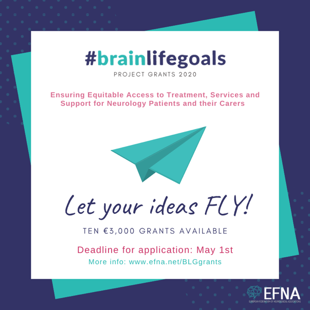
In 2020, within the framework of the #BrainLifeGoals campaign, the European Federation of Neurological Associations (EFNA) will again provide grants to support work in one of their focus areas. This year the focus will be on:
Ensuring Equitable Access to Treatment, Services and Support for Neurology Patients and their Carers.
Donna Walsh, EFNA Executive Director, explained “Access to treatment is one of the central #BrainLifeGoals of many neurology patients, and we want to help make that a reality across Europe! Therefore, EFNA will provide up to 10 grants of €3,000 each to selected organisations.”
The closing date for applications is May 1st.
Find out about eligibility and requirements here: https://www.efna.net/blggrants/

Mar 19, 2020
Stroke and dementia rank among the most pressing health issues in Europe. Cerebral small vessel diseases (SVDs) have emerged as a central link between these two major co-morbidities. SVDs account for more than 30% of strokes and at least 40% of dementia cases. They encounter multiple distinct diseases that can be separated based on their underlying genetic defects, risk factors, and clinical presentations. Despite this profound impact on human health, there are no treatments with proven efficacy against SVDs. The network “Small vessel diseases in a mechanistic perspective: Targets for Intervention in Stroke and Dementia(SVDs@target)” brings together top scientists with a wide range of complementary expertise. We spoke with Danielle Kerkhofs, PhD candidate from the Maastricht University about this project and the latest developments.
SAFE: If you were to explain the project’s aim to a person without any medical background, what would you say?
DK: The SVDs@target project aims to elucidate underlying mechanisms of cerebral Small Vessel Disease (cSVD) and discover new treatment options for this disease. CSVD is an umbrella term used for different pathologies affecting the smallest vessels in the brain. It contributes to a quarter of all strokes and almost 45% of all dementia’s. With revealing the underlying mechanisms of the disease we hope to create possibilities to develop new treatments specific for CSVD.
SAFE: What types of partner do you need to carry out a project like this?
DK: The partners that we need for this project should have both clinical as pre-clinical research experience. To further reveal the underlying mechanism of the disease we need to start at a basic level, followed by clinical studies in patients. I think this balance between the pre-clinical and clinical research is one of the strengths of this project.
SAFE: Can you briefly describe your role in the project?
DK: I am working as a PhD student on this project at Maastricht University, participating both in pre-clinical as clinical studies. Our main research topic in Maastricht is to investigate the specific role of inflammation, and more specific different immune cell populations, in the development of cSVD. Further I participate in the clinical studies Investigate@SVDs and TREAT-SVDs.
SAFE: What personally attracted you to be in this project?
DK: What I really like in this project is the internationally collaboration between the different research groups and the focus on both preclinical as clinical work.
SAFE: When this project ends, what do you expect to change, i.e. how it will reflect on stroke treatment?
DK: This project will give us more insight in the mechanisms underlying the pathogenesis of cSVD. The acquired knowledge will hopefully make the next step possible were we can investigate more specific treatments that can reduce progression of the disease. Further this new knowledge can also provide us new chances for earlier detection of the disease.
SVDs@Target has received funding from the European Union’s Horizon 2020 research and innovation programme under grant agreement No. 666881.
Mar 16, 2020
In the midst of the COVID-19 Pandemic, several European physicians have reported a reallocation of stroke resources and reduction of hospital staff due to quarantine or infection. Francesco Corea, FESO, Chair of the ESO Social Media Committee and Marialuisa Zedde, Chair of the ESO PR Committee, each took some time to record their experiences and observations in Italy, one of the first and hardest hit European countries thus far.
“While the price in terms of victims for COVID19 increases there can be further dramatic repercussions for many other diseases and clinical fields. The toll could be very high. My personal point of view, the Italian health care system suffers the shock of corona virus pandemic. From the news that I have available, several stroke units of large hospitals have been reorganized, moved or even closed to meet the need to assist hundreds of patients with serious infectious and respiratory problems” says Francesco Corea, Ospedale San Giovanni Battista Foligno, Italy. Read the full text here.
“Some practical considerations on how the organization of our daily work has already changed and is still changing in taking care of patients with acute stroke. My point of observation comes from a hospital earlier and more directly involved in the ongoing emergency, being close to Lombardy region. Anyway, it is only a personal view but I hope these experiences and reflections may be useful for other colleagues in different countries” said Marialuisa Zedde, Reggio Emilia Hospital, Italy. You can read her full text here.
We recommend you keep an eye on ESO blog about the Covid19 crisis to stay up to date with the latest developments.
Featured image by Gerd Altmann from Pixabay


Mar 12, 2020
by ESO | 12.3.2020 | ESO |
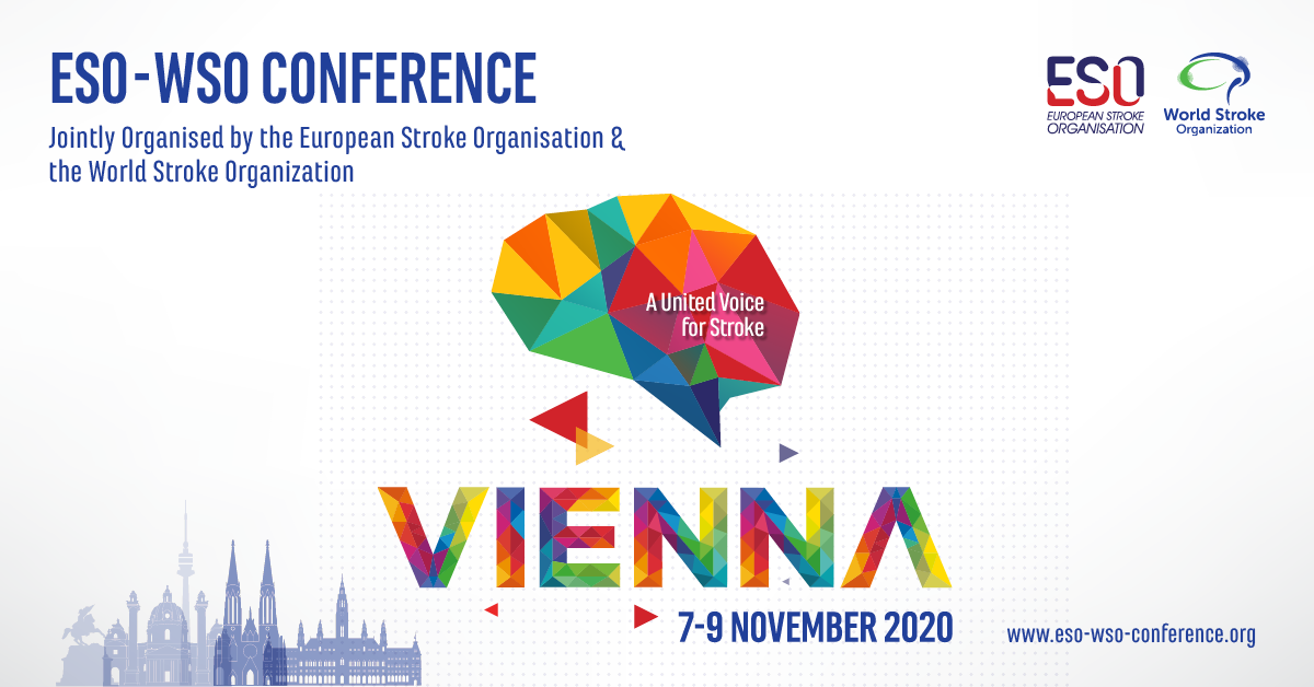
Due to the latest developments regarding the COVID-19 outbreak, and in an effort to prevent spreading the virus and for the sake of event participants, the European Stroke Organisation together with the World Stroke Organization, have decided to postpone the ESO-WSO 2020 Conference, scheduled to take place in Vienna from 12-15 May 2020.
The new dates for ESO-WSO 2020 are 6-9 November. Answers to FAQ related to this postponement can be found on the conference website.
The conference will take place at the same venue, Austria Center Vienna and current registrations will be transferred to the new Conference dates.
In the coming weeks we will be updating the website with new registration deadlines, new programme scheduling and additional information.
Image by ESO.
Mar 9, 2020
First published on ARNI Institute for Stroke Rehabilitation website | March 9, 2020
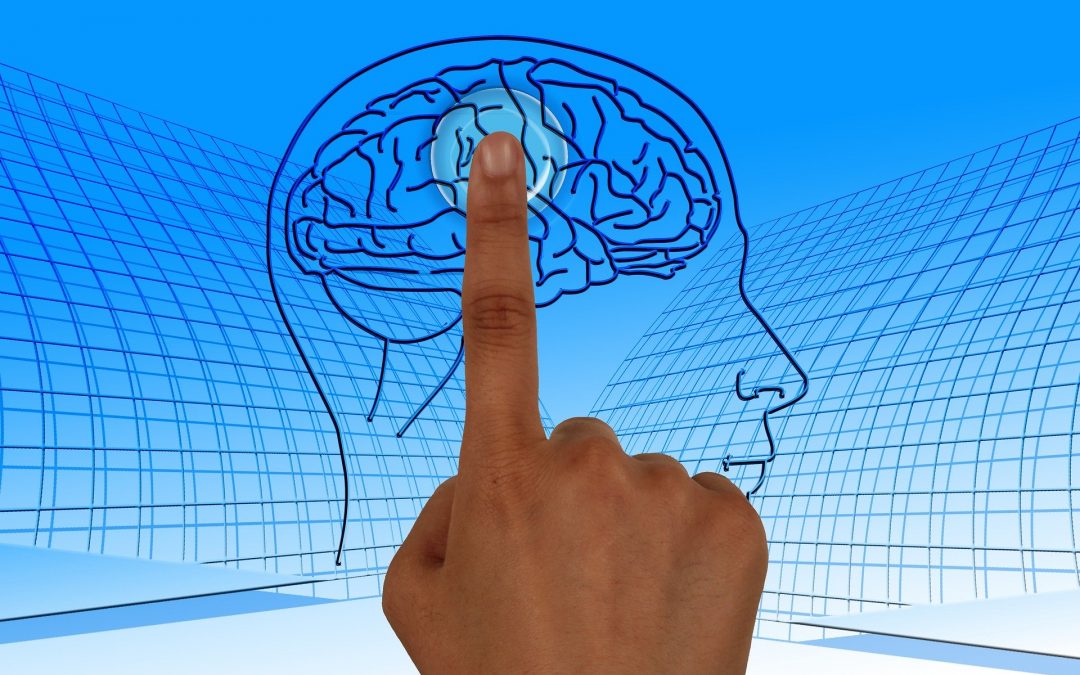
Neuromodulatory non-invasive brain stimulation (NIBS) techniques are experimental therapies for improving motor function after stroke. The aim of neuromodulation is to enhance adaptive or suppress maladaptive processes of post-stroke reorganisation. However, results on the effectiveness of these methods, which include transcranial magnetic stimulation (TMS) and transcranial direct current stimulation (tDCS) are mixed. It’s posited that recent developments in NIBS technology will likely contribute to individualised therapy. Moving beyond single-area stimulation, targeting specific muscle groups that play different roles in post-stroke motor recovery (for example, finger flexors vs. extensors) may well be possible using multi-locus TMS. NIBS in stroke faces a challenge reminiscent of the development of other stroke therapies, such as thrombolysis and mechanical thrombectomy, where early studies were largely mixed before patient selection and individualising protocols were refined to determine its therapeutic potential.
So, researchers at UCL want to find out:
- How brain activity changes after someone has a stroke.
- If weak, non-invasive brain stimulation could encourage the brain into a pattern of brain activity which is useful for upper limb rehabilitation.
Please follow this link to red the full article.
Image by Gerd Altmann from Pixabay
Mar 6, 2020
First published by ESO | 6.3.2020
Author: Dr Nicolas Martinez-Majander, Department of Neurology, Helsinki University Hospital, Finland
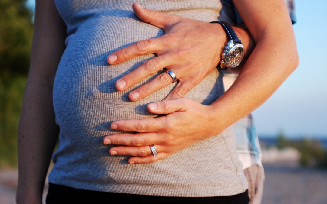
Incidence of young-onset ischemic stroke is currently about 13/100 000 per year in high-income countries and has been increasing since 1980s. In young patients, stroke affects social life, family, and working ability for years after the event. In addition to well-known risk factors, such as diabetes mellitus, hypertension, and hypercholesterolemia, young patients also have unconventional, age-specific risk factors such as pregnancy and puerperium. However, there are still many unanswered questions in terms of risk factors for stroke in the young, and so far study results have been inconsistent.
Ischemic stroke during and after pregnancy
Both pregnancy and puerperium are associated with an increased risk of all stroke subtypes. In a single-center Canadian study, most ischemic strokes occurred during the third trimester, around delivery, or during postnatal period with an incidence of 18 strokes per 100.000 deliveries.(1) Furthermore, of all thromboembolisms during pregnancy, stroke contributed to 12% of them.(2) Up to 25% of ischemic strokes during pregnancy can be associated with eclampsia.(3)Other risk factors for ischemic stroke during pregnancy include e.g. hypertension, diabetes, age over 35 years, black ethnicity, migraine headaches, thrombophilia, smoking, alcohol, and other substance abuse.(4) Kittner et al. also showed that the relative risk of ischemic stroke was up to 8.7 (95% CI, 4.6-16.7) during puerperium.3
The choice of secondary prevention depends mostly on the stroke etiology and gestational age. Aspirin in low daily doses is usually the drug of choice especially during the second and third trimester, but there are inconsistent results regarding its teratogenic effects during the first trimester.5 If anticoagulation is indicated, e.g. in high-risk source of cardioembolism, then low-molecular-weight heparin would be recommended instead of novel anticoagulants or warfarin which crosses placenta and is found to be potentially teratogenic. A few case reports and retrospective studies have shown that in acute treatment, intravenous thrombolysis (rtPA) might be safe during pregnancy, but since these women were excluded from all the randomized clinical trials, pregnancy is still a formal contraindication of rtPA. The same applies to mechanical thrombectomy and treatment decisions should be made on an individual basis for each patient.
You can read the full article and references here.
Illustration: Image by ekseaborn0 from Pixabay
Mar 5, 2020
Every year, around 1.3 million Europeans have a stroke. Twenty to 35% of the patients die in the first month after stroke, and around one third remain dependent on the help of others. The annual costs for stroke care in Europe are estimated at € 64.1 billion. Mainly in the first days after stroke onset, patients are at risk of complications, such as infections and fever. These complications are strongly and independently associated with a higher risk of death or dependency. This week, we bring you an interview with Jeroen de Jonge, Neurology Resident, UMC Utrecht, a trial manager of PRECIOUS, a trial that is looking for ways to improve the recovery of stroke patients aged 66 years or older.
SAFE: If you were to explain the project’s aim to a person without any medical background, what would you say?
The PRECIOUS trial stands for ‘PREvention of Complications to Improve OUtcome in elderly patients with acute Stroke’. This medical trial is an EU-funded Horizon 2020 collaborative research project dedicated to look for ways to improve the recovery of stroke patients aged 66 years or older. After a stroke, patients can have complications, such as fever or a pneumonia. Patients who have a complication after stroke, usually recovery worse than patients without a complication. Normally, the complications are treated when symptoms become obvious. Fever is treated with paracetamol, nausea and vomiting with metoclopramide, and infections are treated with antibiotics. In this trial, we give the treatments before symptoms occur, to test if we can prevent the complications and thereby improve recovery.
SAFE: What types of partner do you need to carry out a project like this?
The setting-up, conduct and management of a trial like PRECIOUS involves many different partners and organizations. PRECIOUS is carried out in about 80 hospitals in 9 European countries. Before starting medical research with patients, it has to be approved by regulatory authorities, which assess if your trial is useful, safe, ethical and feasible. In every participating hospital, there is a dedicated doctor who helps with setting-up the trial. However, sometimes the rules and regulations are so complex, that a specialized research organization is involved to help this process.
During the course of the trial, several partners are involved, each with a specific task. For example, there is a safety team that collects information about potential side effects of the drugs, to keep an eye on the safety of the treatments. A different team designs an electronic data system, which is a safe online platform where local investigators can fill in patient data. Also, there is a team that visits the hospitals periodically, to monitor if the trial is being performed according to the rules. Also, at the end of the trial, a lot of information is gathered, and we need a statistical team to process all the information to determine if the treatments are effective. However, these are just a few examples, and you need a lot of partners to carry out such as project.
SAFE: Can you briefly describe your role in the project?
My role as a trial manager is to oversee all processes related to PRECIOUS on a day-to-day basis. The work is very broad and consists, for example, the general management of the trial, support with obtaining regulatory approvals, setting-up participating hospitals, including patients into the trial, and answering questions of local investigators about patient recruitment, data collection or follow-up of patients.
SAFE: What (if any) are the difficulties with carrying out the work?
Obtaining regulatory approvals in different countries and hospitals requires a lot of paperwork and bureaucratic tasks. This process takes a lot of time. Off course, it is very important that research is assessed very carefully before it can start, but sometimes specific aspects of the research are being assessed multiple times. It takes a lot of time before the research can actually start, and that is sometimes challenging.
SAFE: What personally attracted you to be in this project?
Stroke is the world’s second largest cause of death and the third cause of long-term disability.
Many patients suffer from a stroke on a daily basis. However, there are still limited treatment options available. Because of medical research, there has been considerable progress in the treatment of stroke and new effective treatment options have become available in the past decade. The possibility support research that looks for new ways to treat stroke is what personally attracts me in this project.
SAFE: When this project ends, what do you expect to change, i.e. how it will reflect on stroke treatment?
We hope to find a simple, safe and effective treatment strategy for patients who have had a stroke, which can prevent the development of complications and improve recovery after stroke.
PRECIOUS has received funding from the European Union’s Horizon 2020 research and innovation programme under grant agreement No 634809.
Feb 28, 2020
European Stroke Organisation and World Stroke Organization Conference ESO-WSO 2020
May 12-15, 2020
Austria Center Vienna
Bruno-Kreisky-Platz 1
1220 Vienna, Austria
www.eso-wso-conference.org
The world’s largest and most important congress on the subject of stroke will take place in Austria’s capital from May 12 to 15. For the first time, the European Stroke Organisation (ESO) and the World Stroke Organization (WSO) are jointly organizing this expert conference. Around 6,500 participants are expected, including leading stroke specialists from all over the world. More than 3,000 abstracts have been submitted.
In addition to the initial presentation of large clinical studies, important current topics such as the increasing significance of thrombectomy will be discussed. Another major focus is stroke prevention, which is a global challenge.
For further details on the ESO-WSO 2020, please visit https://eso-wso-conference.org
If you have not yet put a placeholder in your diary for this important event, we encourage you to do so.
Media registration is open – please send an e-mail to reg_esowso20@kenes.com
Press contact
Urban & Schenk medical media consulting
Barbara Urban: +43 664/41 69 4 59, barbara.urban@medical-media-consulting.at
Harald Schenk: +43 664/160 75 99, harald.schenk@medical-media-consulting.at
Yours sincerely,
Prof. Jesse Dawson (ESO) Prof. Michael Brainin (WSO)
Co-Chair of the Conference Planning Group Co-Chair of the Conference Planning Group
Dr. Mira Katan, Prof. Patrik Michel, Prof. Stefan Kiechl, Dr. Vasantha Padma,
Dr. Diana Aquiar de Sousa, Dr. Yvonne Chun (PR Committee / Press Liaison)
Barbara Urban, Harald Schenk (PR Liaison)
Feb 27, 2020
Slovak weekly magazine in cooperation with RTVS -Slovak national TV and Radio and with the Slovak National Theater presented the nominees of the twelfth year of the SLOVENKA OF THE YEAR.
JUDr. Alžbeta Husarovič, MHA, MPH is one of this year’s laureates for the Slovakian Women of the Year. She is the President of the civic association Porážka.sk, which is dedicated to raising stroke awareness and helping patients and their families with information about physiotherapy, speech therapy, psychology, social assistance. The aim of this organisation is to improve patients’ quality of life and lead them to self-sufficiency.
“I was very pleased and amazed when I became the Nominee for the Slovak Woman of the year 2020. I didn’t think that somebody is even noticing me for what I am doing. And I am doing this activity with love” said Alžbeta, adding: “This event Slovak Woman is a competition but I don’t consider it so. For me, it is a opportunity for woman to show others what the can do!”
The Official Press Release “Slovakia 2020”
We have no idea yet of which successful Slovak woman will be awarded in the following categories: Business and Management, Arts and Culture, Media and Communication, Science and Research, Education, Support for Young Talents, Healthcare, Sport and Charity, since all laureates are inspirational ladies, who will be introduced to you over the next few weeks.
In addition, this year the nomination committee will award the title of Absolute Slovak Woman of the Year and the Special Lifetime Achievement Award for Slovakia. We will come to learn the winning names during the gala evening in the historical building of the Slovak National Theater and live broadcast of RTVS – Slovak National TV and Radio on May 31, 2020.
Throwback
Historically, the first Slovak Woman of the year in 2009 became the head physician of the 2nd Pediatric Clinic of the Faculty of Medicine and the Pediatric University Hospital with the Policlinic in Bratislava MUDr. Anna Hlavatá, and so far the last – Slovak of the year 2019 – is Andrea Gontkovičová.
Voting from today at www.slovenkaroka.sk
Starting today, it is possible to vote for individual nominees at www.slovenkaroka.sk and from next week also in the weekly Magazine Slovenka. Vote for your favorite and win an exclusive 8-day vacation stay for 2 people at Labranda Coral Beach 4 * with all inclusive in attractive Gambia from CK Hydrotour.
<End>
Image credits: Porazka.sk


