
Jan 31, 2020
First published on ScenceDaily.com
New research from the Smidt Heart Institute at Cedars-Sinai showed for the first time that women’s blood vessels — including both large and small arteries — age at a faster rate than men’s. The findings, published Wednesday in the journal JAMA Cardiology, could help to explain why women tend to develop different types of cardiovascular disease and with different timing than men.
“Many of us in medicine have long believed that women simply ‘catch up’ to men in terms of their cardiovascular risk,” said Susan Cheng, MD, MPH, MMSc, senior author of the study and director of Public Health Research at the Smidt Heart Institute. “Our research not only confirms that women have different biology and physiology than their male counterparts, but also illustrates why it is that women may be more susceptible to developing certain types of cardiovascular disease and at different points in life.”
Using community-based data amassed from multiple sites across the country, Cheng and her research team conducted sex-specific analyses of measured blood pressure — a critical indicator of cardiovascular risk. The data represented nearly 145,000 blood pressure measurements, collected serially over a 43-year period, from 32,833 study participants ranging in age from 5 to 98 years old.
Because a person’s risk for developing a heart attack, heart failure, or a stroke typically begins with having high blood pressure, Cedars-Sinai researchers combed through their massive data looking for clues and patterns regarding how blood pressure starts to rise. Then, instead of comparing the data from men and women to each other, investigators compared women to women and men to men.
This approach allowed investigators to identify that the progression and evolution of women’s vascular function is very different than for men. In fact, women showed signs of blood pressure elevation much earlier in life than men.
“Our data showed that rates of accelerating blood pressure elevation were significantly higher in women than men, starting earlier in life,” said Cheng, the Erika J. Glazer Chair in Women’s Cardiovascular Health, who also serves as director of Cardiovascular Population Sciences at the Barbra Streisand Women’s Heart Center. “This means that if we define the hypertension threshold the exact same way, a 30-year old woman with high blood pressure is probably at higher risk for cardiovascular disease than a man with high blood pressure at the same age.”
Christine Albert, MD, MPH, founding chair of the newly established Department of Cardiology at the Smidt Heart Institute, says this new research should help guide clinicians and researchers to think differently when it comes to treating and studying women and their cardiovascular health.
You can read the full article here.
Image: Pixabay
Jan 24, 2020
First published on ScienceDaily.com
It’s been almost a quarter century since the first drug was approved for stroke. But what’s even more striking is that only a single drug remains approved today.
In a publication appearing this month in the journal Translational Stroke Research, animal scientists, funded by the National Institutes of Health, present brain-imaging data for a new stroke treatment that supported full recovery in swine, modeled with the same pattern of neurodegeneration as seen in humans with severe stroke.
“It was eye opening and unexpected that you would see such a benefit after having had such a severe stroke,” said Steven Stice, Georgia Research Alliance Eminent Scholar and D.W. Brooks Distinguished Professor in the University of Georgia’s College of Agricultural and Environmental Sciences. “Perhaps the most formidable discovery was that one could recover and do so well after the exosome treatment.”
Stice and his colleagues at UGA’s Regenerative Bioscience Center report the first observational evidence during a midline shift — when the brain is being pushed to one side — to suggest that a minimally invasive and non-operative exosome treatment can now influence the repair and damage that follow a severe stroke.
Exosomes are considered to be powerful mediators of long-distance cell-to-cell communication that can change the behavior of tumor and neighboring cells. The results of the study echo findings from other recent RBC studies using the same licensed exosome technology.
Many patients who suffer stroke exhibit a shift of the brain past its center line — the valley between the left and right part of the brain. Lesions or tumors will induce pressure or inflammation in the brain, causing what typically appears as a straight line to shift.
“Based on results of the exosome treatment in swine, it doesn’t look like lesion volume or the effects of a midline shift matter nearly as much as one would think,” said Franklin West, associate professor of animal and dairy science in the UGA College of Agricultural and Environmental Sciences. “This suggests that, even in some extremely severe cases caused by stroke, you’re still going to recover just as well.”
You can read the full article here.
Image: Pixabay
Jan 24, 2020
First published on EFNA website – Event announcement and invitation
On 18 February 2020, EFNA together with EBC and EAN, will hold the event “Brain Health as a Global Priority – Time for the EU to Act Now” in the European Parliament, Brussels from 11am to 1pm in Room 6Q1, hosted by Jarosław Duda, EPP.
Follow this link to register via EFNA website.
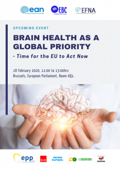 The event is held under the patronage of Members of the European Parliament [EP]:
- Ewa Kopacz, Vice President of the EP, European People’s Party [EPP]
- Mairead McGuinness, Vice President of the EP, European People’s Party [EPP]
- Miriam Dalli, Vice-Chair, Group of the Progressive Alliance of Socialists and Democrats [S&D]
- Frédérique Ries, Vice-Chair, Renew Europe [RE]
- Kateřina Konečná, European United Left–Nordic Green Left [GUE/NGL]
- Tilly Metz, Group of the Greens/European Free Alliance [EFA]
The event looks to explore the following:
Is brain health a global priority?
In the Political Declaration on Non-Communicable Diseases (NCDs), which arose from the United Nations (UN) High-Level Meeting on NCDs in 2018, all Governments recognized that ‘mental disorders and other mental health conditions, as well as neurological disorders, contribute to the global burden of NCDs. It resulted in adding mental and neurological health as the ‘5th NCD’, complementing global efforts to combat cancer, CVD, diabetes and chronic respiratory diseases. This has been reinforced in the recent 2019 UN Declaration on the Universal Health Coverage, where ‘mental disorders and other mental health conditions as well as neurological disorders’ have been identified as an ‘essential component of UHC’. Additionally, the World Health Organisation (WHO) is also now building a new Brain Health team to operate from its headquarters in Geneva. The question now is how can we ensure this progress can be translated into policy action at EU and member state level?
You can access more information and agenda by following this link.

Jan 10, 2020
First published on ScienceDaily.com
Middle-aged adults with high blood pressure, Type 2 diabetes, heart disease or stroke could be at high risk for cancer and early death when sleeping less than six hours per day, according to new research published in the Journal of the American Heart Association, the open access journal of the American Heart Association.
“Our study suggests that achieving normal sleep may be protective for some people with these health conditions and risks,” said lead study author Julio Fernandez-Mendoza, Ph.D., associate professor at Pennsylvania State College of Medicine and sleep psychologist at the Sleep Research & Treatment Center of the Penn State Health Milton S. Hershey Medical Center in Hershey, Pennsylvania. “However, further research is needed to examine whether improving and increasing sleep through medical or behavioral therapies can reduce risk of early death.”
Researchers analyzed data of more than 1,600 adults (20 to 74 years old, more than half women) from the Penn State Adult Cohort who were categorized into two groups as having stage 2 high blood pressure or Type 2 diabetes and having heart disease or stroke. Participants were studied in the sleep laboratory (1991-1998) for one night and then researchers tracked their cause of death up to the end of 2016.
Researchers found:
Of the 512 people who passed away, one-third died of heart disease or stroke and one-fourth died due to cancer.
People who had high blood pressure or diabetes and slept less than 6 hours had twice the increased risk of dying from heart disease or stroke.
People who had heart disease or stroke and slept less than 6 hours had three times the increased risk of dying from cancer.
The increased risk of early death for people with high blood pressure or diabetes was negligible if they slept for more than 6 hours.
You can read the full article here.
Image: Pixabay.com

Jan 7, 2020
First published on ScienceDaily.com
A team of New Jersey stroke researchers has linked recovery of reading and language competence with cerebral blood flow in the left reading network. Their findings may contribute to new approaches to identifying and treating reading deficits after stroke. The open access article, “Cerebral perfusion of the left reading network predicts recovery of reading in subacute to chronic stroke” was epublished on August 26, 2019 in Human Brain Mapping. The authors are Olga Boukrina, PhD, and A.M. Barrett, MD, of Kessler Foundation, and William Graves, PhD, of Rutgers, the State University of New Jersey.
Despite the fundamental role of reading ability in everyday living, little research has been conducted on patterns of reading recovery after stroke, or the development of interventions to improve reading outcomes. In this study of left-brain stroke, a team of New Jersey scientists examined patterns of cerebral perfusion bilaterally, including left and right networks of brain areas important for healthy reading, the area surrounding the stroke lesion, and the corresponding contralateral area.
They enrolled 31 participants during inpatient rehabilitation, within 5 weeks of left-sided stroke. All underwent functional magnetic resonance imaging, psychometric testing, neurological examination and tests for phonological, orthographic and semantic impairments. Fifteen participants had follow-up studies at 3 months post stroke. Analysis of data from the subacute and chronic phases showed that recovery of reading and language competence correlated with increases in cerebral blood flow in the left reading network.
You can read the full article here.
Image: Pixabay.com
Jan 6, 2020
Source: European Journal of Arrhythmia & Electrophysiology. 2019;5(2):77–81
Authors: Kamal Kishor , Devendra Bisht , Sanjay Kalra
Abstract
Atrial fibrillation (AF) is one of the major causes of stroke, heart failure, sudden death and cardiovascular morbidity in the world. Management of AF, its risk factors and complications demands huge cost. The frequent hospitalisations, haemodynamic abnormalities, and thromboembolic events related to AF put a huge emotional and monetary burden on patients, so preventive strategies will be a cost-effective means of reducing this burden. Primordial prevention (i.e., intervention prior to onset of disease risk factors) should be implemented by the government, along with the food industry, to educate the public regarding the importance of a healthy diet and adherence to it. Primary prevention should focus on halting the onset of AF in targeted populations who carry risk factors for development of AF. The strategy at the secondary-prevention level includes optimal control of rate or rhythm either by anti-arrhythmic drugs or catheter ablation. Every effort should be made to reduce the incidence of ischaemic stroke by using optimal oral anticoagulation, devices or surgical closure of left atrial appendage. Tertiary level prevention should focus on the management of complications incurred as a result of AF, such as ischaemic stroke, heart failure or asymptomatic left ventricular dysfunction. Since management of AF demands a huge economic burden and the line between thrombotic complications and bleeding complications is a thin one; over-diagnosis and overtreatment of AF should be avoided. The ultimate aim of quinary prevention is to sensitise the physician to follow evidence-based medicine and to get rid of populated myth. This current review summarises a stepwise preventive strategy for AF from the primordial level to the quinary level.
You can read the full article in the latest exciting issue of European Journal of Arrhythmia & Electrophysiology – Volume 5 Issue 2 by clicking here.
Image: Pixabay.com


