Oct 25, 2020
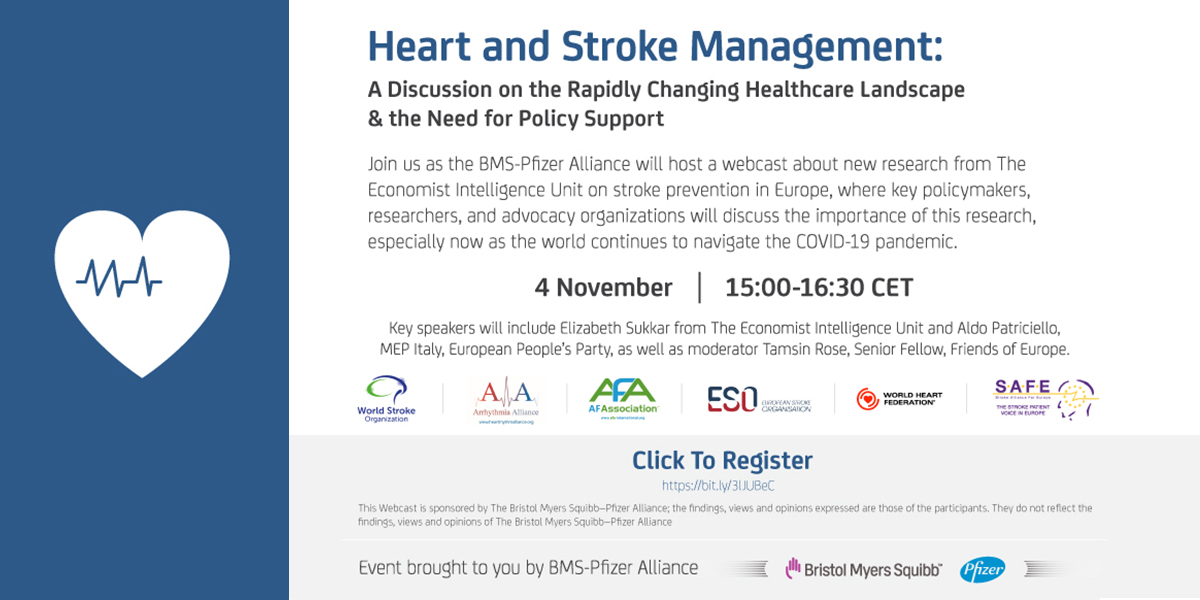
Join us for this webinar: Heart and stroke management: a discussion on the rapidly changing healthcare landscape & the need for policy support
Date: Wednesday, 4 November 2020 Time: 15:00 CEST
Duration: 1 hour, 30 minutes Registration: https://bit.ly/3lJUBeC
Oct 23, 2020
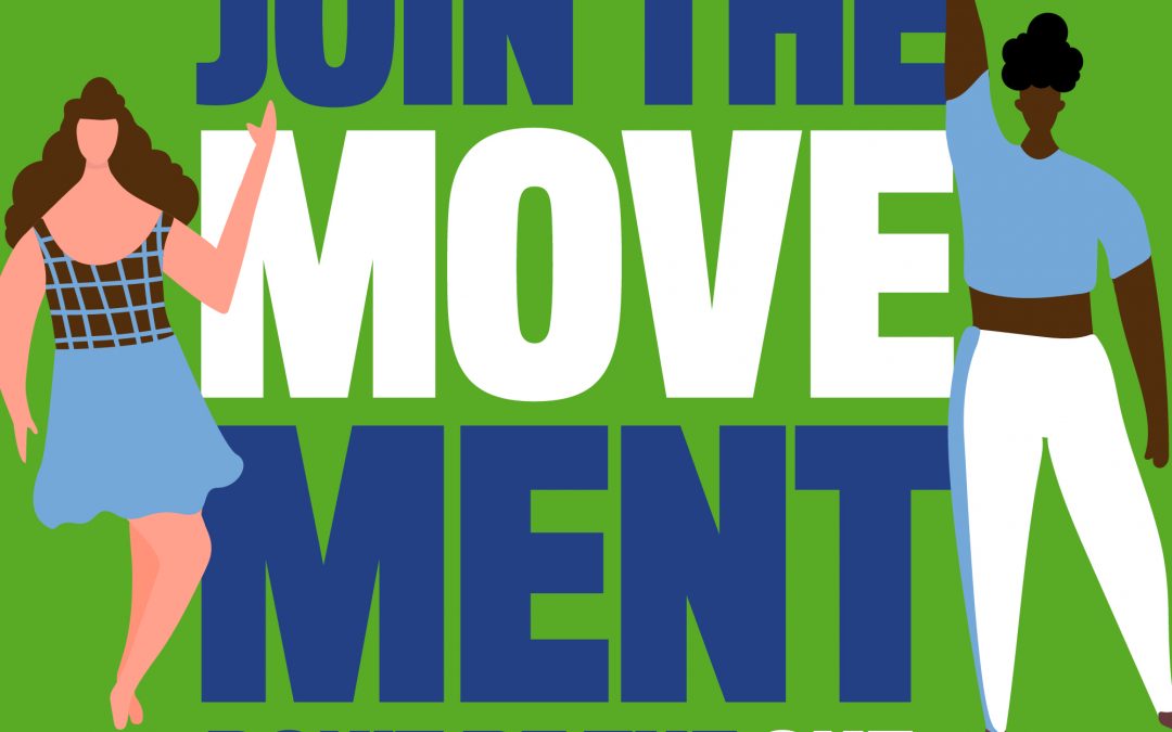
We are proud to support World Stroke Day on 29 October.
1 in 4 of us will have a stroke – Don’t be the One and join the MoveMent – be part of the world’s biggest dance chain to prevent stroke.
Help us raise awareness of stroke on World Stroke Day.
Reach out to everyone you know and ask them to help get the message out and even better to be part of the Global Dance Chain Challenge.
Spread the word about World Stroke Day by sharing on social media and emails.
To find out more follow @WStrokeCampaign or check out their website here
Oct 21, 2020
Dear Friends,
We are in very difficult times. We have never seen a pandemic like this affecting our everyday lives. I hope you and your loved ones are safe and well. We want to let you know that SAFE is here for you and continuing to work despite all the problems around.
While COVID 19 is at the forefront of our thinking, stroke still exists. We know that many of you will be at the front line of services and will be seeing the impact of COVID on individuals who have suffered from stroke. We want to hear from you, we want to know what is happening to stroke survivors in your country, we want to know what is happening to your stroke support organisations and we want to know your ideas as to how we can help.
In addition to finding out how COVID 19 is affecting you and your country, SAFE has adapted its work for 2020. Unfortunately, we have not been able to meet with our members through our usual regional meetings, but we have managed to been up, virtually, June and we will again in November. We have delayed the launch of our economic burden of stroke report until November year. The SAFE General Assembly will be held in November this year and we will do this virtually as we are not able to gather you together for a face to face meeting. I am also excited to let you know that that our first European life after stroke forum is going virtual! The date of 12 March 2021 remains the same and we will share more information with you over the coming months. We will fund one representative from your SSO, and one therapist or nurse from your country. The conference will also be open generally to delegates interested in improving life after stroke services. More information, please click here.
What is happening in the world is a lot for everyone to take in just now, and everyone is having to rapidly adapt their lives on a daily basis. I would like to thank all of you for all the exceptional work you are doing. Please continue to keep well and be safe, do what you can to ensure the survival and sustainability of your stroke support organisation, and we hope to hear from you soon.
With best wishes,
Jon Barrick
President
On behalf of the Board of SAFE

Sep 24, 2020
We are delighted to introduce to two new recruits to SAFE’s team!
Caroline is joining us as our new communications manager and Lora as our new campaigns manager. Please join us in warming welcoming them to the SAFE family.
Click here to find our more!
Sep 18, 2020
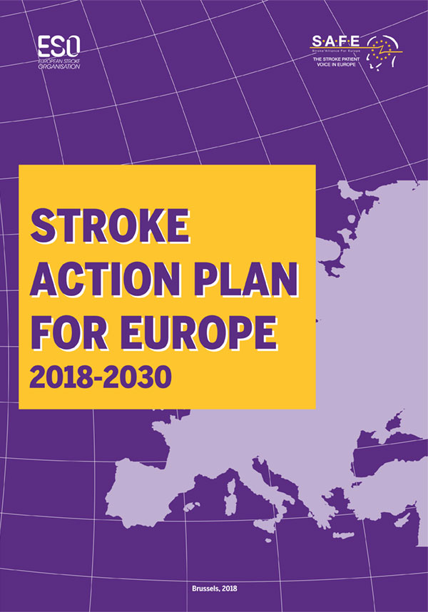
SAFE recently co-hosted the Stroke Action Plan for Europe ‘kick off’ meeting with the European Stroke Organisation. The Plan 2018-2030 outlines a set of key recommendations that if implemented would improve the care and outcomes for stroke patients and stroke survivors across the whole care pathway.
We brought together stroke support organisation and national scientific societies from all over Europe to discuss how the Plan could be implemented across all European countries. This is a very exciting step towards providing better support to stroke patents and stroke survivors. We look forward to sharing more information with you over the coming months.
Here is the press release for more information
Jul 6, 2020
The World Stroke Organsiation is running a three part series titled ‘Maintaining Stroke as a Priority’. In this series stroke support organisations from the Americas, Europe and Asia/Oceania have highlighted the impact of COVID 19 on people with lived experience of stroke across the stroke pathway, discussed the responses and innovations of stroke support organisations and signposted to patient education resources available globally. Join them for the last in their Maintaining Stroke as a Priority webinar series, 14:00 GMT/UK time, 09:00 EDT time, on Wednesday 8 July with:
- Juliet Bouverie (Stroke Association UK): Discharge/transitioning home
- Patrice Lindsay (Heart and Stroke Canada): Secondary prevention
Registration: https://us02web.zoom.us/webinar/register/WN_90ILNiK9Q2SBMbhZNux6Gg


