Dec 18, 2020
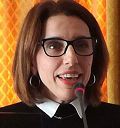
At the SAFE General Assembly last month (November 2020), SAFE members appointed Hariklia Proios as the new President.
Hariklia is has been on the SAFE board for three years and is the Vice President of the Hellenic Action for Stroke in Greece.
We asked Hariklia about her new role and what her ambitions for SAFE are.
Watch the video
Why did you want to be SAFE’s next President?
I truly believe in SAFE’s mission and goals and I am proud to be involved in such an important organisation operating at a global, European level. Having a background in organising Stroke Support Organisations for more than 20 years and having worked on stroke-related rehab both in terms of academic research and also in clinical setups, in the United States, Switzerland and now Greece, I believe I have the professional expertise and personal experience to help lead our joint efforts.
Why do you feel that SAFE’s role is important?
SAFE has been involved in educating, campaigning and encouraging research and support organisations but always from the stroke survivors’ perspective, always engaging all stakeholders, including healthcare professionals and guiding policy- making not only at a European but also at a global level. SAFE helps stroke survivors engage their human stories, lives are disclosed to us and this is one of the privileges of being involved in SAFE. I believe that if we pay attention to each story detail, we learn something in the process about how life exists “before and after stroke” and is full of adaptation and change. Stroke is a life-altering experience and so therefore SAFE’s role of improving care for stroke is of utmost importance. I would like to continue to advocate on behalf of SAFE and behalf of stroke survivors as I strongly believe stroke survivors should have a voice throughout the healthcare pathway.
Do you see SAFE changing direction in the coming years?
SAFE members have different entry points, and this organisation is here to share information on policy work, lobbying and maintaining a patient voice through case study examples which are also included, for example, in the SSOFT online faculty tool. It is clear the future will involve increasingly more virtual meetings even though these meetings were guided by the erratic vicissitudes of today’s pandemic. So, communication modes have changed but SAFE has always continued and will continue its mission and goals. In the future, even when we will return to face-to-face General Assembly meetings, Working Conferences and Country Cluster meetings in member countries, providing information about the expectations of support organisations, best practice and care, we will most likely maintain a significant virtual element of support, thus reaching out to stroke survivors and other stakeholders who cannot physically attend.
What will be your top priority as SAFE’s President?
SAFE has initiated and is leading a number of important projects, the continuation and successful completion of which will be of top priority in the next couple of years. These projects include the Stroke Action Plan (2018-2030), which influences national care in all member countries, iwill guide policy-making at a global level and the Life After Stroke forum (taking place in March 2021) which is capitalising on the knowledge of all engaged stakeholders, healthcare professionals, stroke support organisations, research/academic experts, and energised leaders. These projects are just a few among others which will be a key ingredient for SAFE’s success. SAFE will continue to encourage member countries to promote the Economic Burden of Stroke and will in the future deal methodically with complex stroke issues including regional inequalities (eg. gender and environmental), diversity and gaps, converging in parallel with a clear-cut setting of goal priorities.
What excites you most about your new role as President?
Understanding and supporting each member organisation, empathetically and always with positive regard and through dialogue, will help us find common solutions and get all those affected by stroke the access to the help and support they need. Teamwork transforms visions to sustainable solutions. My dedication is and will remain to strategically develop a sustainable SAFE as well as continue to honour its cultural legacy for wanting to make a difference in people’s lives.
What is your ambition for SAFE?
We realise that we have a big role in instituting knowledge and education by raising expectations of what quality of stroke care should be like, by strengthening stroke survivor organisations. Stroke prevention is feasible, acceptable, and cost effective: monitoring an irregular heartbeat (AF) or early treatments such as getting to the emergency room in due time. We must stay mindful of best practices and making a difference for those lives who cannot help themselves. My ambition is to look for and try to embrace all our members’ interests and together we will continue to increase public awareness about stroke as a strategic priority.
Watch the video
Dec 18, 2020
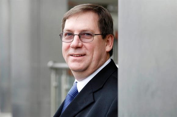
Last month (November 2020), after six years on the board, Jon Barrick stepped down as SAFE’s President and retired from the board.
Jon dedicated many years to being a champion of improving the live of people affected by stroke and we warmly thanked him for this at our recent General Assembly.
Before he left, we asked him about his time with SAFE, and any advice he’d give to the new President, Harriet Proios.
What have you found most rewarding about being President of SAFE?
I’ve done two roles in the SAFE Board, six years as the Secretary and six years as the President. I’ve always been hopeful that I could make a difference to the quality of life of stroke survivors, and to improving the possibilities of surviving a stroke.
Being a part of SAFE was very rewarding because both my parents were stroke victims and SAFE has given me the opportunity to make that difference. More than that I found myself in an organisation that had other people who also wanted to make a difference, and so I felt I was not alone and I had the basis on which progress could be made. I’ve met some truly inspirational people through and in SAFE, many of whom I now count as lifetime friends.
I’ve always been an internationalist and it’s been so rewarding to work in SAFE to help the growth of Stroke Support Organisations throughout Europe. I have enjoyed trying to understand the systems and cultures of different countries, but mainly meeting so many different people has been great. I’ve also found it rewarding to find that people in positions of power in the EU and commercial organisations have been so supportive towards SAFE and its goals, justifying the hope that change for the better can be achieved.
Working for SAFE has also been rewarding because we have had to learn and grow and overcome challenges all the time, it has been hard work at times but with good people working together I’ve learnt that a way to succeed can usually be found. It’s rewarding to see people grow and develop, and to see obstacles overcome. Collective success is more rewarding to me than individual success.
Watch the video
Jon, what was your proudest moment during your time as President?
So many proud moments, but being around people who are building stroke support organisations in their countries always makes me feel humble and proud of their courage and determination. Achieving funding every year makes me proud because it means SAFE survives for another year and can pay its way, for both staff and the projects.
And what great projects we have done, pivotally important projects, such as:
- SSOFT, enabling people to understand about Stroke support Organisations and be able to campaign,
- the Burden of Stroke in Europe the first comprehensive look at stroke numbers now and in the future and make commentary on what needs to change,
- the Economic Impact of Stroke in Europe, again the first study in this area and the platform for showing stroke is an investible proposition
- the campaigning meetings held inside the European Parliament raising the profile with the EU Health commission and MEP’s
- our engagement with Stroke researchers and encouragement at EU level for more research funding
- the development of the Stroke Action Plan for Europe, and patient literature to be available in stroke units, the list goes on….
But probably I’m proudest of the growth of SAFE and its number of members, and the growth in competence of SAFE over this time which means it is set up well for the future.
Watch the video
Jon, what advice would you give to the new President?
Harriet the new President is an excellent colleague, who I’m sure will do a good job. Everyone has to approach the Presidents role to reflect the way they are as a person, and so enthusiasm, good interpersonal skills, and knowledge of stroke will all be a natural part of the new Presidents make-up. My advice would be to remember that enabling stroke support organisations in their countries is the key component of SAFE’s work, from that comes the survivability of SAFE as well as strength at country level across Europe.
I think two streams of work will have to dominate the next 10 years, firstly the development and successful running of the European Life after Stroke Forum, which should aim to be the largest gathering of its sort in the world, and can be that if it’s driven forward with passion. SAFE can secure both its financial stability, but also drive forward quality of life through this initiative. Secondly the pursuit of the Stroke Action Plan for Europe, which will have benefits for all campaigners in the field of stroke, at EU and national level, is very central and something to which other projects can feed into.
Finally enjoy it, enjoy being with the Board and the team, keep everyone informed and involved, to be President of SAFE is a privilege given to few.
Watch the video

Nov 13, 2020
The 27th November is the deadline for the abstract submissions for the 1st European Life After Stroke Forum.
We are calling on for applications in two areas:
- Scientific applications: this may be for completed for ongoing trials and studies in the broad area of life after stroke.
- Grab and Steal: this is to share experiences of service developments in life after stroke where these original ideas and innovative practices could be used by others.
We are seeking abstracts which may give those interested an opportunity to present either a ten-minute oral presentation or a poster presentation.
For more information on how to apply – click here: https://bit.ly/31xwCIS
Please share with your contacts – we are really looking forward to receiving excellent applications, whether scientifically, or service development focused, from all over Europe.
Nov 13, 2020
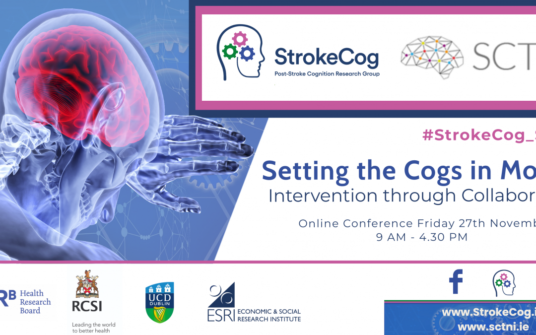
Our President, Jon Barrick, will be presenting at ‘Setting the Cogs in Motion: Intervention though Collaboration’ conference on 27th November 2020.
The event focuses on post-stroke cognitive impairment and promises to be a great day of exciting talks and presentations from speakers including Professor Terry Quinn, University of Glasgow and Professor Urs Fischer, University Hospital, Bern, members of the StrokeCog team, representatives from Headway Ireland, Irish Heart Foundation, Family Careers Ireland and The Stroke Alliance for Europe, among many others.
There will be a mix of patient/public, researcher, policy and health-care professional speakers and this is set to be a very interesting event!
Registration is FREE, but places are limited so register early!
Link to website – – www.strokecog.ie/conference
Link to Registration – https://bit.ly/3mMuWTc
Join the conversation on Twitter @StrokeCog and @SCTNIreland by using the official hashtag #StrokeCog_SCTNI
Nov 9, 2020
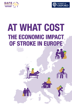
Brussels, 9 November 2020 – New research published today shows future costs of stroke care in Europe could increase to €86 billion in 2040 if we fail to invest in stroke prevention, treatments and rehabilitation.
The study was commissioned by SAFE and undertaken by the heath economics team at the University of Oxford, pre COVID-19.
At What Cost – the Economic Impact of Stroke in Europe [add in link] adds to the existing evidence that shows stroke care is woefully underfunded and needs urgent investment.
Lead researcher, Dr Ramon Luengo-Fernandez says: “The research was completed before COVID-19. Since the beginning of the pandemic, services have been diverted away from non-infectious conditions, including stroke care, therefore we feel that the projected costs in our report are likely to be even higher.”
The study included all stroke-related costs that occurred in the EU countries plus the UK, Israel, Norway, Iceland and Switzerland in 2017.
Report findings show for the first time the cost of stroke along the whole stroke care pathway, including not only direct healthcare costs but also the costs of informal care and productivity losses due to disability or death from stroke. The study provides scientific projections related to stroke costs for the next 20 years.
In addition, the report looks at three interventions which are in the latest stroke guidelines – the treatment of atrial fibrillation to prevent stroke, mechanical thrombectomy (the clot retrieval from the blood vessel in the brain) in the acute phase of stroke and community-based rehabilitation after stroke. The research shows that no matter at which point in the stroke pathway you intervene – prevention, treatment, long-term care – there’s likely big gains to be had in terms of patient outcomes and cost savings.
In 2017, nearly 1.5 million people suffered a stroke, nine million Europeans lived with a stroke, and more than 430,000 people died due to a stroke in the 32 countries under this study. The total cost of stroke in 2017 was a €60 billion.
The number of new strokes and the number of people living with stroke is set to rise due to ageing population in Europe, as age is the biggest, non-modifiable risk factor for stroke. The costs of stroke are projected to increase by 44% between 2017 and 2040, with some countries seeing rises in stroke-related costs of nearly 100%.
SAFE’S Director General, Arlene Wilkie adds, “Health systems across Europe have been left crushed and broken by COVID-19. Stroke is preventable and it is crucial that we invest in care and support that can respond to the current and future needs of stroke survivors.
This research offers solutions to help reduce the burden of stroke and future proof healthcare services at the time when COVID-19 has amplified the problem and exposed already overstretched and failing healthcare systems. SAFE calls on the EU and national governments to make stroke care a priority and ensure it is fit for the future.”
Summary of findings report
Full research report
Oct 26, 2020
We are excited to announce the first ever European Life After Stroke Forum is going virtual.
With so much uncertainty in the midst of the COVID-19 pandemic, we can now ensure that this important event will be able to go ahead and be accessible to many more delegates across Europe.
If the ELASF is not already in your calendar, make sure you add 12 March 2021 and watch this space for more information in the next few weeks.
We’ll be will post regular updates on our website, including how to register, on our website and on social media, in the coming months. If you would like to know more – why not follow us on Twitter @StrokeEurope #ELASF or sign up to our enewsletter for regular updates
Research and investment in all aspects of the life after stroke care pathway has been neglected. This forum is unique and crucial in bringing together the latest research and needs of stroke survivors to contribute to the SAPE and ultimately improve life after stroke across Europe.
Image by Alexandra_Koch from Pixabay


