Nov 17, 2017
Having worked for the Stroke Association in the UK for ten years, I had a pretty good understanding of the differences in stroke care across Europe. But it wasn’t until I starting editing SAFE’s Burden of Stroke report earlier this year, that the stark inequalities became clear.
It is outrageous to me that your chance of dying from a stroke varies so much, simply due to where you live. As well as causing heartache to tens of thousands of people, not providing good stroke care is a heavy burden on society as a whole. And what good is expensive research into the best stroke prevention, treatment and support, if the findings are not implemented? (more…)
Nov 14, 2017
If you have a heart attack or stroke, it’s important to get your “bad” cholesterol measured by your doctor on a follow up visit. Researchers have found that one step is significantly associated with a reduced risk of suffering another serious cardiovascular episode.
The new research, conducted by researchers at the Intermountain Medical Center Heart Institute in Salt Lake City, found that patients who don’t follow up with their doctor by getting a low-density lipoprotein (LDL) cholesterol test following a heart attack or stroke are significantly more likely to have a reccurrence. (more…)
Nov 14, 2017
Patients with a prior history of heart attacks or stroke have better outcomes when cholesterol-lowering medications are used after they’re discharged from the hospital, according to a new study from the Intermountain Medical Center Heart Institute in Salt Lake City.
Prior surveys in hospitals found that statins, a common medication prescribed to lower cholesterol, aren’t being used as consistently in patients who’ve been admitted to the hospital following a heart attack or stroke. Researchers also found that when the medication is prescribed, dosing is likely not as high as it should be to provide optimal benefits. (more…)
Nov 14, 2017
Standard guidelines for stroke treatment currently recommend clot removal only within six hours of stroke onset. But a milestone study with results published in the New England Journal of Medicine shows that clot removal up to 24 hours after stroke led to significantly reduced disability for properly selected patients.
The international multi-center clinical study, known as the DAWN trial, randomly assigned 206 stroke victims who arrived at the hospital within six to 24 hours to either endovascular clot removal therapy, known as thrombectomy, or to standard medical therapy. (more…)
Nov 12, 2017
Picture a bare wire, without its regular plastic coating. It’s exposed to the elements and risks being degraded. And, without insulation, it may not conduct electricity as well as a coated wire. Now, imagine this wire is inside your brain.
That’s what happens in many diseases of the nervous system, such as multiple sclerosis (MS), spinal cord injuries, stroke, neonatal brain injuries, and even Alzheimer’s disease.
Much like that bare wire, the nerve fibers in the brain lose their protective coating, called myelin, and become extremely vulnerable. This leaves the nerve cells exposed to their environment and reduces their ability to transmit signals quickly, resulting in impaired cognition, sensation, and movement. (more…)

Nov 9, 2017
Bo Norrving is a professor in neurology at Lund University, Sweden. He has authored more than 400 publications on clinical stroke research, including several seminal papers e.g. the Swedish Aspirin Low Dose Study, and the world’s largest study of stroke in the young. He is a founder of the Swedish Stroke Register (Riksstroke), the world’s 1st national stroke registry. He is a member of the advisory group for ICD 11 at WHO. He was the President of the World Stroke Organization (WSO) 2008-2012, and chairs the WSO Global Policy Committee. He is the editor-in-chief of the European Stroke Journal.
Having in mind that he was one of the editors of the important 2006 Helsingborg Declaration on European stroke strategies, the European Stroke Organisation and SAFE are proud and honored to have Prof. Norrving now coordinating a joint ESO and SAFE project called “Stroke Action Plan for Europe 2018-2030”.

Prof. Bo Norrving; Photo: Henrik Rosenqvist
SAFE: Stroke Support Organisations are only just beginning to be formed and to grow in quite a lot of countries; do you think that medics are fully aware of how important they are in influencing decision makers on allocations of funding and resources to things like stroke care, stroke research and public awareness? If not as aware what is ESO doing to encourage medics to aid the growth of SSO?
BN: I think the full importance is still not well recognized. There are many examples where the initiatives of SSOs have been critically important. It’s an important task for ESO to support formation of new SSOs and to support and collaborate with existing ones. ESO and SAFE have recently joined forces in several actions and projects (e.g. at the EU, the European Stroke Action Plan), and I am sure there will be many more in the future. (more…)

Nov 3, 2017
A new and exciting e-learning tool is being developed by SAFE to support Stroke Support Organisations around positive and effective stroke advocacy
The Stroke Alliance For Europe (SAFE) in partnership with the European Stroke Organisation (ESO) has begun to develop a new online e-learning tool for empowering stroke advocates from across Europe. The Stroke Support Organisation Faculty Tool (SSOFT) will help to build the capacity and capabilities of Stroke Support Organisations (SSOs) by developing their knowledge and understanding around how to build positive and effective advoacy campaigns for stroke prevention, diagnosis and treatment. (more…)
Nov 3, 2017
The 11th World Stroke Congress promises to attract acclaimed experts in stroke from around the world. The congress will showcase a cutting-edge educational and scientific experience, focusing on the latest developments in stroke prevention, acute management and restorative care after stroke.
This major Congress will be returning to North America for the first time in more than 12 years.
Canada has well-developed stroke care systems and has contributed to significant advances in stroke care through efforts in developing guidelines, clinical and health systems research. These efforts are ongoing and increasingly important in the face of an aging population and increased health care demands in Canada and around the world. (more…)
Nov 2, 2017
Researchers at Maastricht University Medical Center and Maastricht University have discovered why the brain is more sensitive to oxygen deprivation, or hypoxia, than other organs. Hypoxia caused by a stroke, for example, activates a specific mechanism that is protective in other organs but can be detrimental to the brain. ‘This discovery solves a long-standing mystery of the unique sensitivity of the brain to hypoxia,’ says head researcher and professor Harald Schmidt. The research results were published today in the leading scientific journal Proceedings of the National Academy of Sciences (PNAS). (more…)
Nov 1, 2017
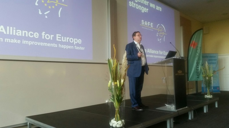
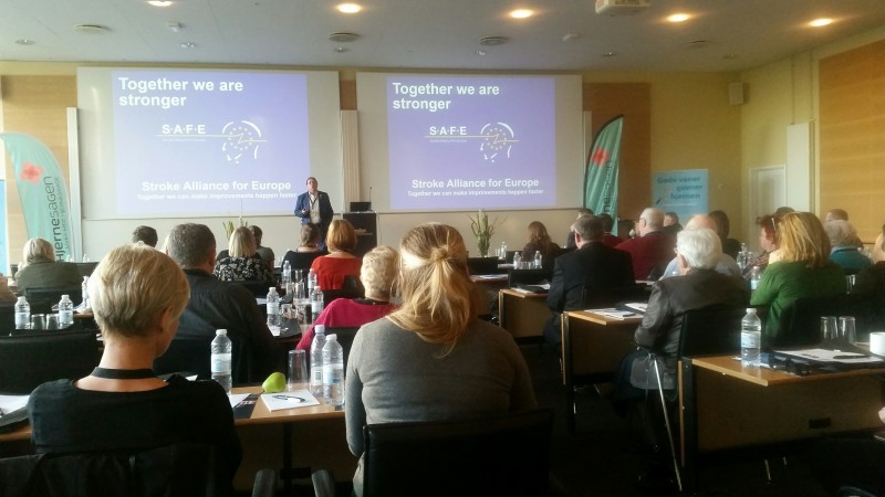
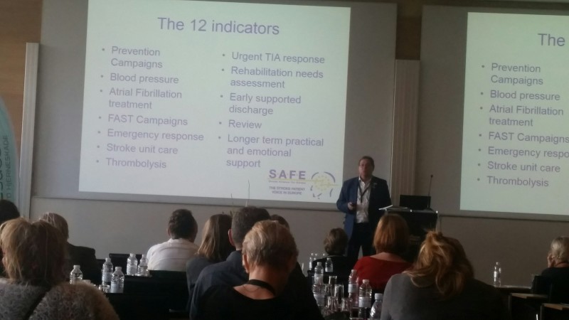
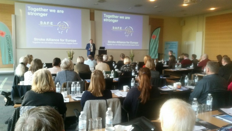
Research needs to be the platform for action in the decision making and policy sphere- said Jon Barrick, SAFE President at the World Stroke Day event in Denmark, on 30th October 2017.
Please see here the full presentation from this event.