Feb 7, 2019
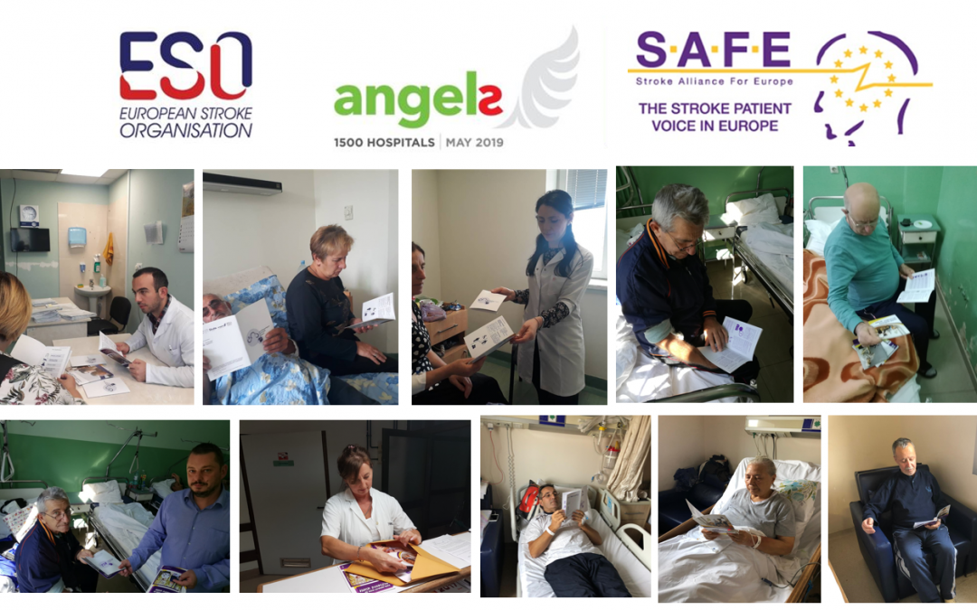
The Angels Initiative is a unique healthcare initiative launched by Boehringer Ingelheim in partnership with the European Stroke Organisation (ESO), the World Stroke Organisation (WSO) and the Stroke Alliance for Europe (SAFE).
SAFE’s members proudly contributed to the Angles Initiative by providing critical information to stroke patients and their families as early as in stroke units. In addition to containing useful and evidence-based information, these brochures help improve patient – doctor communication as well.
The SAFE Angels project was realised in the first half of 2018. It involved translation to 13 European languages, design, printing and distribution of five different brochures for stroke patients and their carers. The core texts were kindly provided by Stroke Association UK.
13 organisations from the following countries took part in this project and made sure these brochures were made available in the stroke units: Spain, Serbia, Poland, Czech Republic, Latvia, Croatia, Macedonia, Greece, Ukraine, Georgia, Hungary and Turkey.
Patient information brochures are available only in a selected number of hospitals in the project countries, but can also be downloaded from SAFE website.
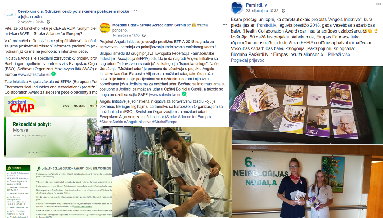
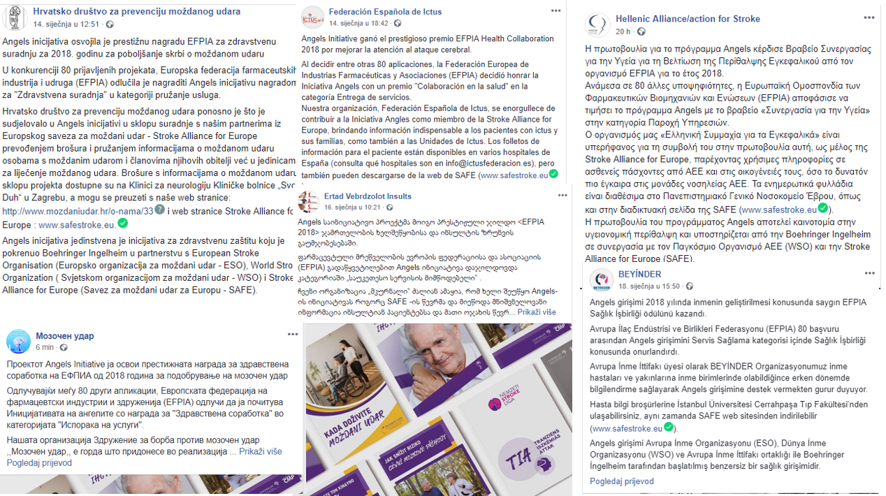
“We consider this as a pilot project, because we already distributed the whole quantity of the patient information brochures and now both patients and medical staff are asking when we will have it again. For now, we are signposting people to visit SAFE’s website and download the PDF versions.”
Ivan Milojevic, Serbian stroke support organisation Mozdani udar
In 2018, the Angels Initiative won the prestigious 2018 EFPIA Health Collaboration Award for Improving Stroke Care.
Deciding among 80 other applications, the European Federation of Pharmaceutical Industries and Associations (EFPIA) decided to honor the Angels Initiative with a “Health Collaboration” award in the category Service Delivery.
SAFE members proudly reported about this event, reminding their audiences once again of when these brochures can still be found in 12 European countries. For more detailed information, please see our Facebook page or look up for a specific stroke support organisation’s Facebook profile.

Feb 5, 2019
This year’s first SAFE Board meeting took place in London this week, from 4-5th February.
The Board members took two days to discuss the current and future SAFE Strategy, with special focus on governance, finances and supported growth in member organisations.
Among many SAFE ongoing projects, there will be two which are very important for SAFE’s political activities. The first and the fast approaching is the EU Presidency event that is going to organised in the EU Parliament on 28th March this year. The Board discussed the details around this project and the activities that will be performed prior to the event to ensure better impact. The other strategically important project is the big research about the Economic Impact of Stroke in Europe. This research is being done by the Oxford University researchers and the report is to be presented in the EU Parliament at the end of October this year, around the World Stroke Day.
The Board ratified the Social Media Strategy and agreed on ways forward and the venues for this year’s Regional Conferences were defined.
Stroke Support Organisation Faculty Tool (SSOFT) was again among the most important topics, considering its value and significance for the stroke support organisations’ empowerment.
Jan 31, 2019
This article first appeared on ARNI Stroke Charity (UK) website | Author: Tom Balchin
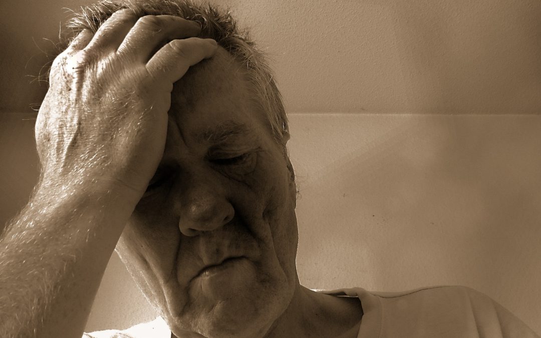
Tiredness is something we all experience in our everyday lives. But fatigue is where we experience tiredness which is unrelated to physical or mental exertion, and is not alleviated by rest. Up to 70% of survivors experience fatigue, characterised by overwhelming physical and/or mental tiredness or exhaustion. For many the symptoms dissipate and lessen over time. Others continue to experience these symptoms at a high level many years after their stroke. This is called chronic fatigue.
It is a condition which can greatly impact upon the quality of an individual’s life, making everyday tasks feel overwhelming and unachievable, or just plain exhausting.
Previously, it was thought that patients who experience depression post-stroke were fatigued as a result of their mental health, whereas it is now highly possible that the inverse relationship may, in fact, be true. Fatigue may often be the cause, or a significant contributing factor, of depression.
There is currently no clinical method for diagnosing fatigue, and no treatment is available to alleviate the condition.
Research into fatigue is at its very early stages. Work to contribute towards a treatment has now been spearheaded by Dr Anna Kuppuswamy, the lead researcher on the project.
Her study aims to further general understanding of how fatigue works in the brain, and whether or not it can be alleviated. The goal for the future is to be able to diagnose and treat fatigue effectively, so that no-one need experience its debilitating effects.
So, how can you help?
STROKE SURVIVORS EXPERIENCING HIGH LEVELS OF FATIGUE –
Please come to help Dr Kuppuswamy’s Team test a new intervention for fatigue!
You can read the full article here.
For more information about the UK ARNI Institute please visit their website.
Jan 24, 2019
The article first appeared on Irish Heart Foundation website | Author: June Shannon
Air pollution and climate change, those who refuse vaccines and antibiotic resistance also listed among the top ten threats to global health in 2019 by the WHO
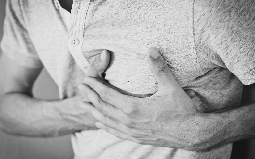
The World Health Organisation (WHO) has identified non-communicable diseases which include cardiovascular disease (heart disease and stroke), as one of the top ten threats to global health in 2019.
A noncommunicable disease (NCD) is a chronic medical condition or disease that is not infectious and cannot be transmitted among people.
According to the WHO, non-communicable diseases, such as diabetes, cancer and heart disease, are collectively responsible for more than 70 per cent of all deaths worldwide, or 41 million people. This includes 15 million people dying prematurely, between the ages of 30 and 69.
Non-communicable diseases, such as diabetes, cancer and heart disease, are collectively responsible for more than 70 per cent of all deaths worldwide or 41 million people.
Non-communicable diseases, such as diabetes, cancer and heart disease, are collectively responsible for more than 70 per cent of all deaths worldwide or 41 million people.
“Over 85 per cent of these premature deaths are in low- and middle-income countries. The rise of these diseases has been driven by five major risk factors: tobacco use, physical inactivity, the harmful use of alcohol, unhealthy diets and air pollution. These risk factors also exacerbate mental health issues, that may originate from an early age: half of all mental illness begins by the age of 14, but most cases go undetected and untreated – suicide is the second leading cause of death among 15-19-year-olds,” the WHO stated.
The WHO added that it would work with governments to help them meet the global target of reducing physical inactivity by 15 per cent by 2030.
In 2016 more than 9,000 people in Ireland lost their lives to cardiovascular disease with almost half dying from heart disease.
Cardiovascular disease includes all diseases of the heart and circulation but most commonly it refers to coronary heart disease (angina, heart attack), stroke and other blood vessel diseases. Other conditions include congenital heart disease, heart valve disease and disease of the heart muscle (cardiomyopathy).
Read the full article here.
Jan 18, 2019
Europe is ageing and stroke is (still) predominantly happening to older people. Cerebral small vessel disease, a condition affecting small blood vessels in our brains is often called ‘the most common aging brain problem that you may have never heard of’.
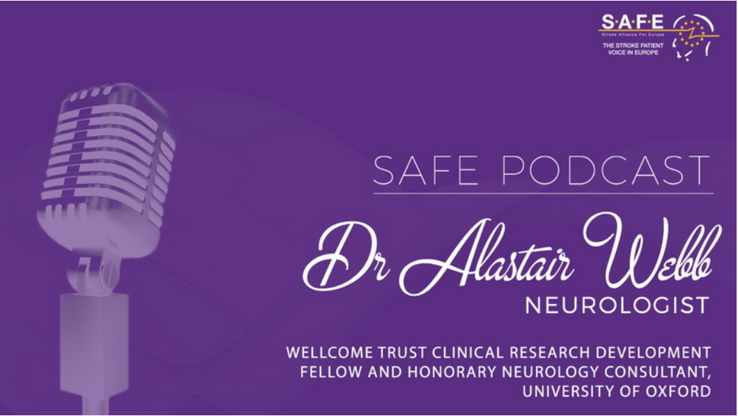
However, it is related to stroke (SVDs account for more than 30% of strokes) and it would be beneficial for people to know more about it. SAFE spoke with Dr Alastair Webb, neurologist from University of Oxford about this condition and what is the relation between SVDs, high blood pressure and stroke.