Sep 5, 2021
Register NOW for FREE for the Life after stroke: caring for the caregiver webinar: bit.ly/3xucXqx
At this meeting we will address some of the challenging aspects posed to informal carers and the impact that caring for a stroke survivor might have on their lives. It will also cover predictors of wellbeing and resilience in carers and good practice examples of carer support and how to implement them.
Find out more about this event here: www.eslaf.org
Sep 5, 2021
The Action Statement on Heart Failure launched on 7 September 2021 outlines policy recommendations and urges the EU to recognise heart failure as a health priority.
The statement supports growing calls for the development of an EU Action Plan on Cardiovascular Disease, similar to Europe’s Beating Cancer Plan. Click here to find out more.
Aug 26, 2021
We are delighted to let you know that Professor Carlo Semenza from Italy and Dr Nuno Ferreira, Professor of Clinical Health Psychology at the University of Nicosia will be presenting at the Life After Stroke: caring for the caregiver webinar on 29 September 2021.
They will be joined by stroke support organisation Cerebrum, Czech Republic, who will be sharing their experiences of developing support for informal carers.
Book your free place now at bit.ly/3xucXqx
Aug 17, 2021
The European Stroke Organisation is holding their the 7th Conference (ESOC 2021) at the beginning of September.
It will be a virtual conference and you register here: www.eso-conference.org
Aug 17, 2021
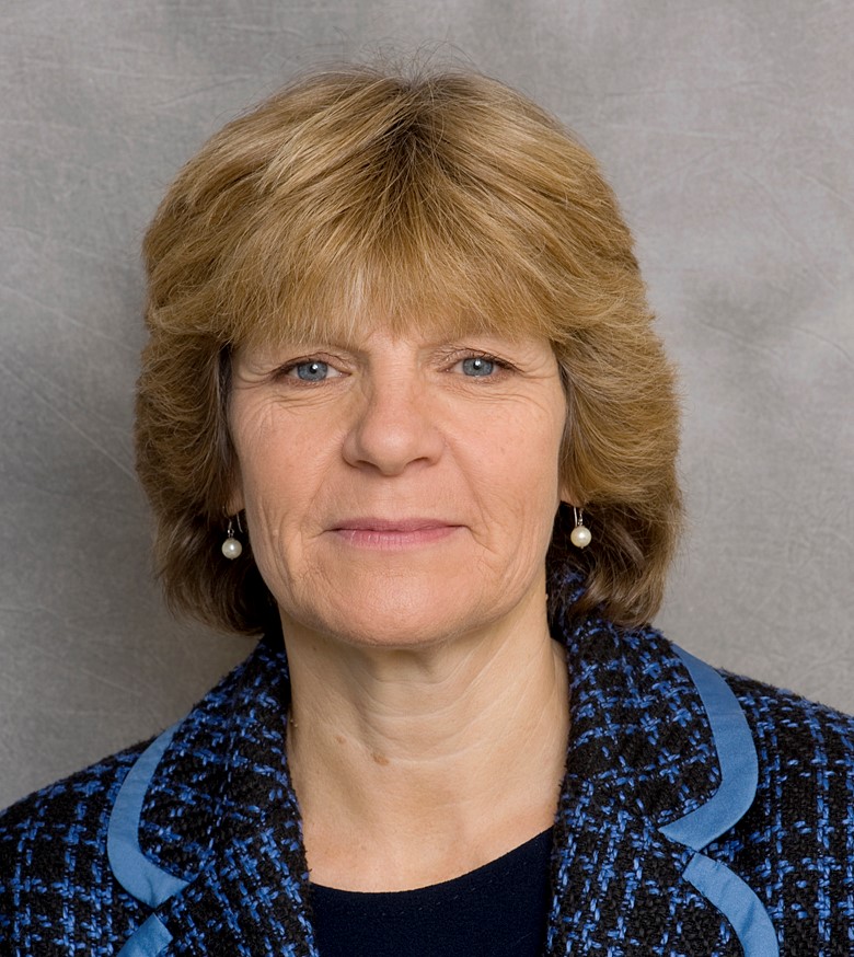
Chair of the European Life After Stroke Forum Scientific Committee, Professor Avril Drummond, highlights the lack of emphasis on improving life after stroke for survivors and how this must be addressed.
‘ the needs of stroke survivors and their families….has not previously been given the focus it deserves’
‘few opportunities exist….in this neglected but vital area of care’
The European Life After Stroke Forum series of webinars in 2021 aims to build the life after stroke healthcare, research and patient community, stimulate debate and ultimately improve life after stroke care and build interest for our 1st European Life After Stroke Forum on 11 March 2022. To book your place on the next free webinar on Caring for the caregiver go to https://bit.ly/3xucXqx
Download the pdf here
For more information on the European Life After Stroke webinars go to https://www.elasf.org/
Aug 11, 2021
The European Brain Council has updated its factsheet on stroke and now includes more information on rehabilitation and life after stroke.
The Stroke factsheet is one of several disease specific factsheets and part of the #ILoveMyBrain initiative.
Find out more at braincouncil.eu/projects/ilovemybrain/


